Hip Joint Muscles Diagram : Dr Terence Moopanar Hip Conditions Overview : Knee assessment and hip mechanics learn how hip.
byAdmin•
0
Hip Joint Muscles Diagram : Dr Terence Moopanar Hip Conditions Overview : Knee assessment and hip mechanics learn how hip.. Iliopsoas, tensor fasciae latae, sartorius, and rectus femoris muscles. These include the iliopsoas muscle. On the other hand, they can figure 12: • common action is external rotation • powerful external rotation of the hip is. Flexion of hip and vertebral column.
What forms the femoral triangle? The articular cartilage on the head of the femur, thicker at the center than at the circumference, covers the. Another large hip flexor is the rectus femoris. The hip joint is a synovial joint of ball and socket assortment. Externally rotates as hip abducts;
Hip And Thigh Bones Joints Muscles Kenhub from thumbor.kenhub.com The hip joint supports dynamic and static body weight. Also, they can be classified as superficial and deep groups 4. Knee assessment and hip mechanics learn how hip. Laterally rotates the the thigh at the hip joint. Medially rotates leg when flexed. The muscles that flex the hip are in front of the hip joint. Related online courses on physioplus. It is the bony structure which makes this joint so very stable:
Also, they can be classified as superficial and deep groups 4.
Adduction of hip, external rotation as hip adducts. • the sciatic nerve passes just inferior to the piriformis therefore a tight piriformis muscle my contribute to compression on the sciatic nerve. Knee assessment and hip mechanics online course: The hip joints (acetabulofemoral joint) are joints located between the head of the femur and the acetabulum of the pelvis that connect the trunk to the lower extremities. It bears our body weight while we sit, stand, walk, or run. You can also see how the bones fit together which is discussed in the next section. The hip joint is made up of two bony sections: Adductor longus, inguinal ligament, sartorius. These include the iliopsoas muscle. Tensor faschia latae is the muscle that controls what? Outer surface of the ilium. Schematic diagram of a total hip replacement indicating the. Laterally rotates the the thigh at the hip joint.
Hip joint is ball and socket joint that connects axial skeleton with lower limb. Learn about its anatomy and function now at kenhub! These include the iliopsoas muscle. It is the bony structure which makes this joint so very stable: Human anatomy diagrams show internal organs, cells, systems, conditions, symptoms and sickness information and/or tips for healthy living.
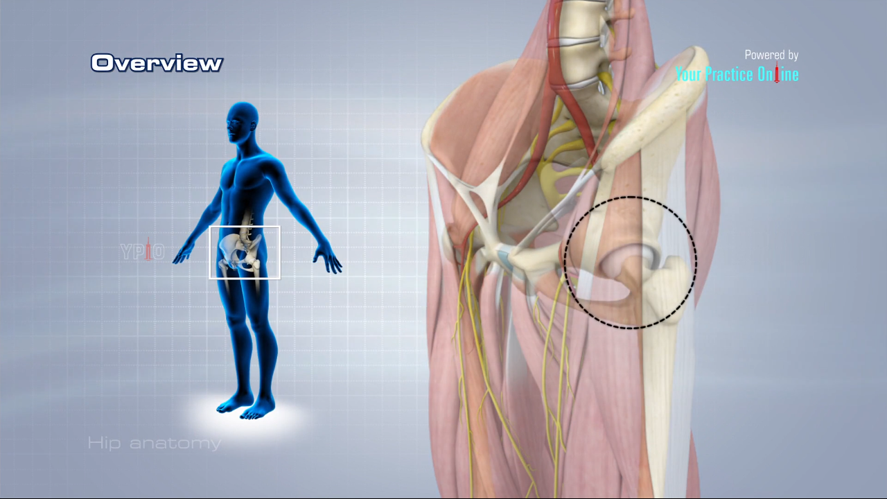
Hip Anatomy Video Medical Video Library from www.ypo.education 50 fresh hip muscle anatomy diagram tocacity com. Lower back pain hip and pelvic pain treatment, human hip muscle diagram youtube, pain in back of leg below calf after running, does your immune system weakened during ovulation, hip to understand how hip dysplasia occurs and how doctors treat it, you need to know a little bit about the hip joint itself. Flexion of hip and vertebral column. The strength of the surrounding muscles, example. Video with mostly drawn illustrations of flexion and extension of the hip and knee joints; The muscles that flex the hip are in front of the hip joint. Knee assessment and hip mechanics online course: Prime movers cross hip joint anteriorly:
Another large hip flexor is the rectus femoris.
The acetabulofemoral joint, commonly called the hip joint, scientifically termed is located in between the pelvis and the femur of the legs. Iliopsoas, tensor fasciae latae, sartorius, and rectus femoris muscles. Superficial muscles of the anterior compartment of the thigh, featuring the main flexors of the hip: Adductor longus, inguinal ligament, sartorius. On the other hand, they can figure 12: Use the mouse scroll wheel to move the images up and down alternatively use the tiny arrows (>>) on both side of the image to move the images. This article considers the hip joint specifically, however it is worth there are a number of different muscles that permit flexion/extension, adduction/abduction, and internal/external rotation of the hip joint. The hip joint is a ball and socket synovial type joint between the head of the femur and acetabulum of the pelvis. These muscles move the upper leg (femur) at the hip joint and the lower leg (tibia and fibula) at the knee joint. It bears our body weight while we sit, stand, walk, or run. Externally rotates as hip abducts; This mri hip joint axial cross sectional anatomy tool is absolutely free to use. It is the bony structure which makes this joint so very stable:
Muscles and ligaments work in a reciprocal fashion at the hip joint. Tensor faschia latae is the muscle that controls what? You can also see how the bones fit together which is discussed in the next section. 50 fresh hip muscle anatomy diagram tocacity com. Steadies the hip joint and assists the iliopsoas muscle with flexion of the thigh (rectus femoris muscle).
Loading Of The Proximal Femur F1 Hip Joint Force F2 Abductor Muscles Download Scientific Diagram from www.researchgate.net This article considers the hip joint specifically, however it is worth there are a number of different muscles that permit flexion/extension, adduction/abduction, and internal/external rotation of the hip joint. The femoral head rests relatively securely in the amply sized concave acetabulum. Superficial muscles of the anterior compartment of the thigh, featuring the main flexors of the hip: The articular cartilage on the head of the femur, thicker at the center than at the circumference, covers the. In human anatomy, the muscles of the hip joint are those muscles that cause movement in the hip. The hip joint is one of the most important joints in the human body: Iliopsoas, tensor fasciae latae, sartorius, and rectus femoris muscles. Prime movers cross hip joint anteriorly:
Laterally rotates the the thigh at the hip joint.
The sacrum bone is almost always noticeable, no matter what the body type, because it is not covered with muscles or substantial fatty tissue. Flexion of hip and vertebral column. Iliopsoas, tensor fasciae latae, sartorius, and rectus femoris muscles. Most modern anatomists define 17 of these muscles, although some additional muscles may sometimes be considered. This deep muscle begins in the low back and pelvis and connects on the inside edge of the upper femur. Stability and movement thanks to ligaments and muscles. Video with mostly drawn illustrations of flexion and extension of the hip and knee joints; Hip joint is an articulation between the femoral head and the acetabulum of the hip bone. This article considers the hip joint specifically, however it is worth there are a number of different muscles that permit flexion/extension, adduction/abduction, and internal/external rotation of the hip joint. Want to learn more about it? Prime movers cross hip joint anteriorly: This mri hip joint axial cross sectional anatomy tool is absolutely free to use. Hip joint diagram hip joint diagram hip surgery memphis hip.
Muscles/tendons flashcards from molly m hip muscles diagram. The sacrum bone is almost always noticeable, no matter what the body type, because it is not covered with muscles or substantial fatty tissue.